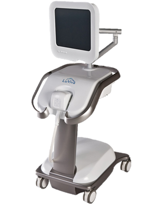
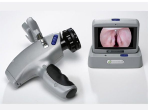
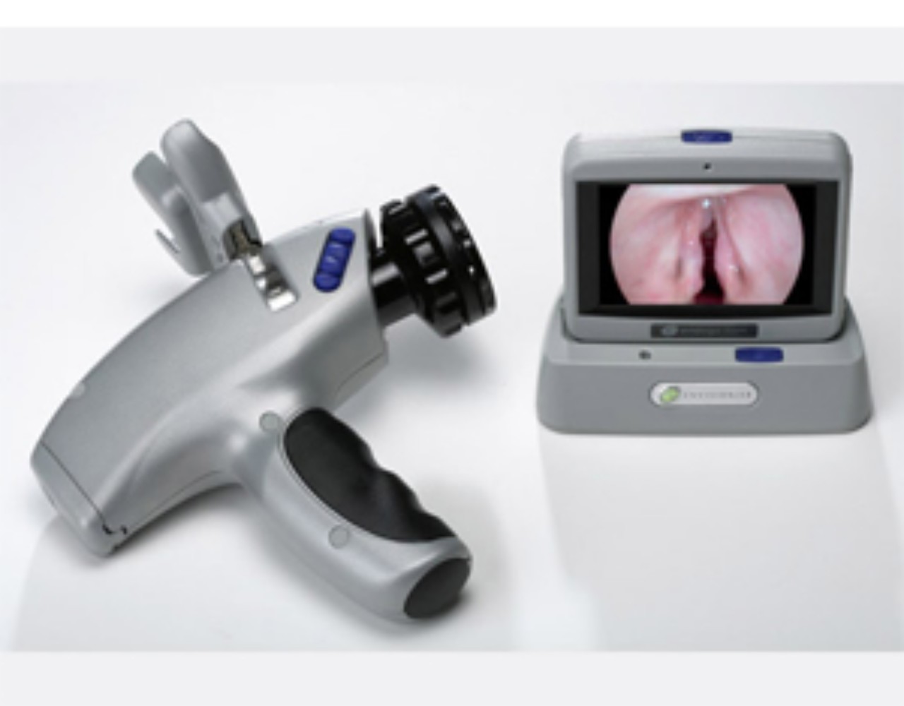
LuViva® Advanced Cervical Scan
Detected Disease up 2 Years Earlier Than Cytologies, HPV Tests, Pap Smear an Biopsies No Need Pap Smear or HPV; No Need Pathology
The LuViva Advanced Cervical Scan is a medical device that has been designed based on biophotonics science. Biophotonics is used in medicine to study tissue and blood at the macro (large-scale) and micro (very small scale) level to detect, diagnose and treat diseases in ways that are non-invasive to the body.
LuViva is based on 18 technology patents developed by the US based company, Guided Therapeutics, Inc. The technology combines fluorescence with reflectance spectroscopy and analyzes very fine gradations of light from tissue. This form of biophotonics is called Multimodal Hyperspectroscopy (MHS).
The purpose of MHS technology is to eliminate disadvantages of the conventional screening processes (high ratio of false negatives / false positives, unnecessary anxiety and concern, unnecessary colposcopy/biopsy procedures, unnecessary surgeries..etc) and provide a more effective and successful cervical cancer screening process.
LuViva can detect physical and chemical changes in the tissue that occur at the cellular level through reflectance and fluorescence spectroscopy. Thus, high grade cellular dysplasia (HSIL) can be recognised by a far higher sensitivity of 87% compared to conventional methods. Because the MHS technology used by LuViva is very sensitive, it may detect early cellular changes starting at the deep basal layer up to 2 years before conventional methods are able to.During a LuViva scan, light from a Xenon lamp is shined upon the cervix through a hollow tube called the Cervical Guide. Using unique algorithms, the interactions of the light with the cervical tissue are analyzed in order to determine the likelihood of precancerous or cancerous tissue being present.
In addition, a short time after certain molecules in the tissue absorb the light, electrons are excited which then cause the molecule to emit light in longer wavelenghths, a phenomenon called fluorescence. The amount of light absorbed by the tissue and the intensity of light reflected and emitted, along with the structure of the emission spectra, depend on the metabolism and structures within the tissue.
The responses to light and the emission of healthy tissue are different than the responses and emission of unhealthy tissue because of the physical changes (increased number of cells, changes in the nuclear size, thickening of the cellular wall..etc) and chemical changes (increased levels of Tryptophan, collagen, elastin …etc) that occur when cells are becoming cancerous.
What is MHS?
Spectroscopy has been a very reliable method used for precise measurements in astronomy and chemistry for a long time. Though there are many types of spectroscopy, all forms function the same fundamental way by using a spectrometer to measure how light interacts with matter. In Multimodal Hyperspectroscopy (MHS), more than one type of spectroscopy is used to analyze very large amounts of spectral data. When these large amounts of spectral information are analyzed, it is called „hyperspectral“. By using a Xenon light bulb to produce the specific waves of light needed, LuViva can scan the cervix in about one minute, with results displayed right away. As the light is applied to the tissue, most is absorbed or scattered, but a portion is reflected by the tissue to the spectrometer. This is called reflectance spectroscopy.


